What skin diseases are precancerous and how do they look?
Read Time:6 Minute, 18 Second
Precancerous– In accordance with the statistics of oncological morbidity in Russia, cutaneous malignant neoplasms among men and women occupy the second place. Over the past ten years, their number has grown by about 30%. The average incidence among women is 13.7%, among men – about 9.9%.
The concept of precancerous skin diseases
Despite the fact that the malignant process of the skin is in visual accessibility, nevertheless, the staginess and variability of its development, as well as the wide variety of benign dermatological pathologies, often do not allow timely detection of a cancer tumor. In connection with the foregoing, the problem of early diagnosis and treatment of a “painful” disease or pathological condition, that is, a precancer has acquired great relevance in dermatology and oncology, against which a development of malignant neoplasm is more or less likely. Read more: Learn to control anger with these simple tricks
Precancerous skin diseases are multiple or single papules, nodules, growths, spots, foci of hyperkeratosis or irritation of various forms, color and size, etc. They are benign epithelial formations and pathological conditions of non-tumor origin, but which can be transformed into malignant tumors. Read more: How to Forget. I work the free tool that facilitates the process to erase your past in Google and Bing
The cause of malignancy can be:
various external factors of a non-specific nature – frequent or persistent mechanical irritation or trauma, prolonged exposure to chemicals (aniline dyes, derivatives of oil, coal, arsenic, pesticides), exposure to temperature factors (frostbite and burns), excessive sun exposure, ionizing radiation, weathering;
endogenous factors (in the body itself) – endocrine disorders, disorders of the immune system and some others;
the absence of timely treatment of benign pathology;
age – among people of middle and old age, transformation into cancer occurs much more often.
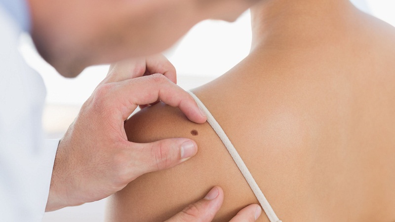
Diagnosis and treatment of precancerous skin pathologies are performed by specialists of a dermatological or oncological profile.
Classification and diagnostics
The generally accepted classification of precancerous pathologies of the skin is still not developed due to the lack of sufficiently clear theoretical notions about these pathological conditions. Therefore, the main ones are very conditionally divided into 2 groups:
Obligatory precancerous skin diseases
Obligatory pathological conditions are characterized by a high probability of transformation into the malignant formation. These include:
- pigment xeroderma;
- limited precancerous hyperkeratosis of the skin of the red border of the lips;
- Bowen’s disease;
- Kery’s erythroplasia;
- Paget’s disease.
Three recent pathologies are currently considered as a special form of skin cancer (cancer in situ – in situ), which is a local pre-invasive (intraepithelial) malignant process that does not go beyond the skin. However, they are traditionally referred to as precancerous pathological conditions.
Pigmented xeroderma
It is genetically caused by a staged disease, which is characterized by excessive sensitivity to even insignificant solar radiation. The first symptoms of this pathology with the defeat of the skin of the face and hands appear already in the first 3 years of the child’s life. After a brief stay in the sun in the open areas of skin, there are limited erythematous spots and small pigmentary yellowish-brownish patches rising above the skin surface, which resemble freckles. The number of them gradually increases, the skin in these places becomes dry and flaky. Later, atrophic changes develop in their place, areas with telangiectasias and a smooth shiny surface appear. Still later, ulcers and cracks, papillomatous and warty growths occur on this background, Fertilization occurs in 100% of cases. Malignant formations have a high tendency to disintegrate and metastasize. Most patients die before the age of 15 to 20 years due to the generalization of the tumor process. However, although the gene of this pathology occurs in approximately 0.28% of people, it is inherited in an autosomal recessive type, and the disease develops only in one of 250,000 cases. Treatment of pigmentary xeroderma is carried out after histological examination. With single sprouting, it consists of electroexcision, laser or cryodestruction, with multiple – in the course of close-focus X-ray therapy.
Limited precancerous hyperkeratosis of the skin of the red border of the lips
Unlike other obligate precancerous diseases, it often affects people of young and middle age. Among the whole of the precancerous pathology of the lips, 80% fall precisely on this form of the disease. The lesion of polygonal shape and sizes from 2 mm to 1.5 cm is localized, mainly on the lower lip directly on the red border. It is surrounded by a whitish thin roller, located approximately in the middle between the corner of the mouth and the center of the lip, soft and painful on palpation. The level of its surface in most patients is slightly below the level of the surrounding red lip rim, because of what the lesion seems sunk. The surface is covered with grayish-brownish dense scales. Usually, education exists for several years in a benign condition, but sometimes it is transformed into a malignant tumor already within the first year and even the first months from the moment of its appearance. The main symptoms that partially help in the diagnosis of malignancy in education are:
- the appearance of a seal in its base;
- the appearance of erosion on its surface;
- strengthening the processes of keratinization.
Treatment consists of electroexcision, laser destruction or surgical excision within healthy tissues, followed by a histological examination. The outcome of the latter depends on the decision of the question of further treatment.
Bowen’s disease
It occurs with the same frequency among men and women over 40 years of age. The factors provoking the development of precancerous state are ultraviolet rays and some toxic substances (arsenic, tar, tar). Any parts of the skin can be affected, but most often – closed (trunk, genital organs), less often – the face and neck.
Optional precancerous skin diseases
The diseases of this group are combined with a relatively low degree of probability of degeneration into cancer. These include mainly:
actinic, or solar, senile (senile) keratosis;
cutaneous horn;
keratoacanthoma;
Actinic keratosis
Develops, as a rule, in the mature and elderly age in people with fair skin in exposed areas that are exposed to long-term (no less than 10-20 years) sun radiation. The basis of the disease is the dysplastic processes of the epithelium, as a result of which it is able to degenerate into squamous cell carcinoma. Usually, senile keratosis is localized to the skin (in the region of the back of the nose, cheeks, ear shells, scalp, lower lip, and hands) that has changed (with uneven pigmentation, thinned) sun. Depending on the severity of clinical symptoms, stages or types of lesions are distinguished:
Erythematous, which is the beginning of the disease and is characterized by the appearance of reddish-pink plaques and spots with sharply delineated boundaries. The diameter of the elements initially does not exceed a few millimeters, and then gradually increases to 1-2 cm. The spots and plaques may have irregular, oval or rounded shape and a rough, rough surface, against which background “telescoping” of telangiectasias is noted and bleeding occurs after easy scraping.
Conclusion
Precancerous-a group of dermatological diseases, against which can develop malignant tumors. Conditionally divided into two subgroups: optional (with optional malignancy) and obligate (with a high likelihood of malignancy). Are single or multiple nodules, proliferation, foci of hyperkeratosis, papules, pigment spots or foci of irritation of different color, size, and shape. The diagnosis is made on the basis of the examination and the results of the histological examination. Treatment – surgical removal, cryotherapy, chemotherapy, interferon therapy.
It may like: http://www.speakymagazine.com/thermal-insulation/
Happy
0 %
Sad
0 %
Excited
0 %
Sleepy
0 %
Angry
0 %
Surprise
0 %


Average Rating